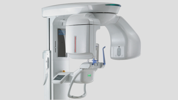
A vital instrument in the precise diagnosis of many head and neck and orofacial conditions is radiographic imaging. 3D imaging is a more elaborate method of obtaining this information and provides a clearer picture as to what exactly pertains to a patient’s condition beneath the surface.
Cone-beam computed tomography (CT) is the emerging choice for 3D imaging and is gradually becoming the standard of care for many dental specialties. Similar to conventional CT scans, sections of an area of the body are taken for relative comparison. However, cone-beam CT differs in that cone-shaped sections are used. These give a better representation of structures relative to surrounding tissues and spaces. It further reduces the number of radiographs a patient has to take, as it provides all the information required in a few sections. This, together with the relative low dose of radiation, reduces the duration of radiation exposure for a patient.
It also aids in 3D reconstruction of images and thus is perfect for demonstrating images of facial profiles before and after treatment. As this method of imaging allows for extensive digital manipulation, it can also be used for more concise orthognathic and orthodontic assessment and treatment planning, among other things. Images can be magnified, superimposing tissues may be filtered out, and even the contrast of the image can be adjusted to produce a more representative image of what is being examined/imaged. Open- and closed-mouth images can be taken with cone-beam CT to accurately examine joint structures and help diagnose orofacial and temporomandibular joint disorder (TMJ) pain syndromes, among others.
It has extensive application in implantology, oral and maxillofacial surgery, and orthodontics as well as many other dental specialties.
A larger area of the facial region, including the eyes, neck and spinal structures, can be viewed with the cone-beam CT. This detail in cone-beam CTs can help in the early detection of metastatic lesions, sialoliths, degenerative diseases and pathologic fractures, which may be missed, especially when asymptomatic. This helps for a more holistic assessment of the dental patient.
One cone-beam CT is representative of all baseline radiographs that may be required for any one patient. It is therefore a safer and more efficient and cost-effective way of accurate clinical diagnosis.
Source: Linda J. Golden, DDS, of Golden Dental Wellness Center (444 Community Dr., Ste. 204, Manhasset). For more information or to schedule an appointment, call 516-627-8400.


